Digital twins in the health sector: mirage or reality?
Digital twins, which are already well established in industry, are becoming increasingly present in the health sector. There is a wide range of potential applications for both diagnosis and treatment, but the technology is mostly still in the research phase.
The health sector is currently undergoing digital transition with a view to developing “4P” treatment: personalized, predictive, preventive and participative. Digital simulation is a key tool in these changes. It consists in creating a model of an object, process or physical process on which different hypotheses can be tested. Today, patients are treated when they fall sick based on clinical studies of tens or, at best, thousands of other people, with no real consideration of the individual’s personal characteristics. Will each person one day have their own digital twin to allow prediction of the development of acute or chronic diseases based on their genetic profile and environmental factors, and anticipation of their response to different treatments?
“There are a lots of hopes and dreams built on this idea,” admits Stéphane Avril, Researcher at Mines Saint-Étienne. “The profession of doctor or surgeon is currently practiced as an art on the strength of experience acquired over time. The idea is that a program could combine the experience of a thousand doctors, but in reality there is a huge amount of work still to do to create equations for and integrate non-mathematical knowledge and skills in very different fields such as immunology and the cardio-vascular system.” We are still a long way from simulating an entire human being. “But in certain fields, digital twins provide excellent predictions that are even better than those of a practitioner,” adds Stéphane Avril.
From imaging to the operating room
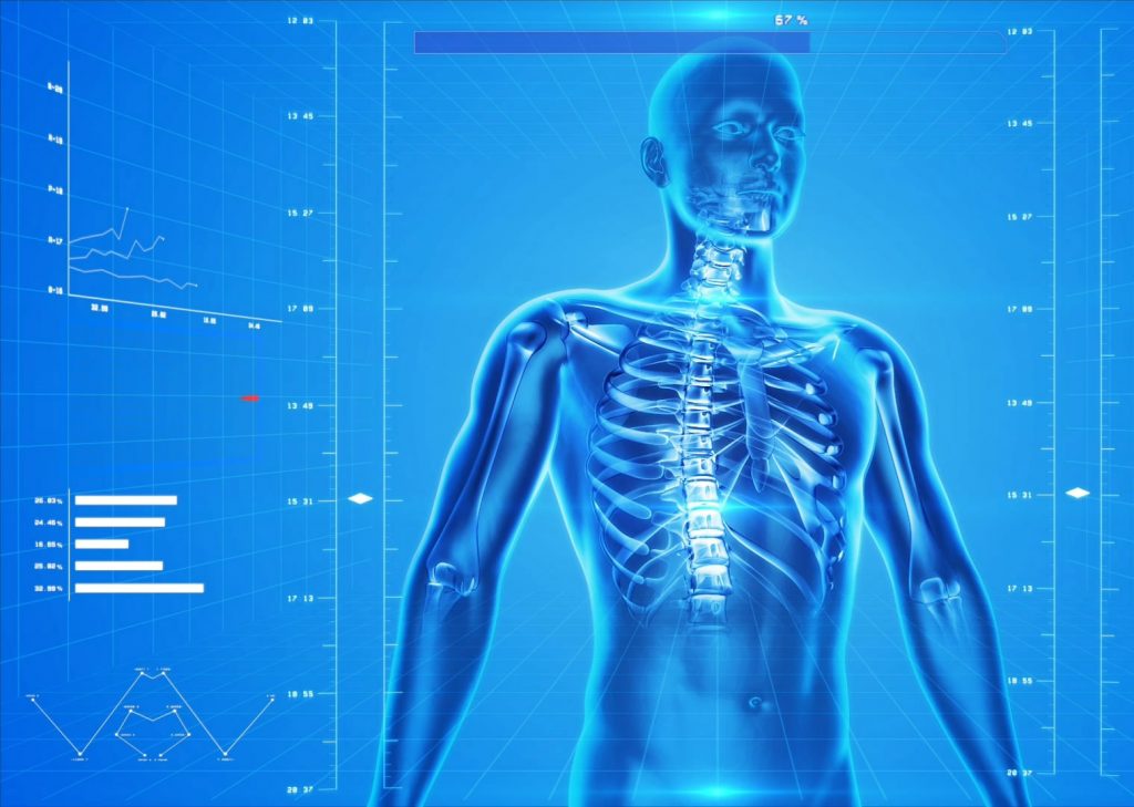
Biomechanics, for example, is a field of research that lends itself very well to digital simulation. Stéphane Avril’s research addresses aortic aneurysms. 3D digital twins of the affected area are developed based on medical images. The approach has led to the creation of a start-up called Predisurge, whose software allows the creation of individual endoprostheses. “The FDA [American Food and Drug Administration] is encouraging the use of digital simulation for validating the market entry of prostheses, both orthopedic and vascular,” explains the researcher from St Etienne.
Digital simulation also helps surgeons prepare for operations because the software provides predictions on the insertion and effects of these endoprostheses once in place, as well as simulating any pre-surgical complications that could arise. “This technique is currently still in the testing and validation phase, but it could have a very promising impact on reducing surgery time and complications,” stresses Stéphane Avril. The team at Mines Saint-Étienne is currently working on improving our understanding of the properties of the aortic wall using four-dimensional MRI and mechanical tests on aneurysm tissue removed during the insertion of prostheses. The idea is to validate a digital twin designed using 4D MRI images, which could predict the future rupture or stability of an aneurysm and indicate the need for surgery or not.
Read more on I’MTech: A digital twin of the aorta to prevent Aneurysm rupture
Catalin Fetita, a researcher at Télécom SudParis, also uses digital simulation in the field of imaging, but this time in the case of air-borne transmission alongside pulmonary parenchyma analysis. The aim of her work is to obtain biomarkers from medical images for a more precise definition of pathological phenomena in respiratory diseases such as asthma, chronic obstructive pulmonary disease (COPD) and idiopathic interstitial pneumonia (IIP). The model allows assessment of an organ’s functioning based on its morphology. “Digital simulation is used to identify the type of dysfunction and its exact location, quantify it, predict changes in the disease and optimize the treatment process,” explains Catalin Fetita.
Ethical and technical barriers
The problem of data security and anonymity is currently at the heart of ethical and legal debates. For the moment, researchers are having great difficulty accessing databases to “feed” their programs. “To obtain medical images, we have to establish a relationship of trust with a hospital radiologist, get them interested in our work and involve them in the project. We need images to be precisely labeled for the model to be relevant.” Especially since the analysis of medical images can vary from one radiologist to another. “Ideally, we would like to have access to a database of images that have been analyzed by a panel of experts with a consensus on their interpretation,” Catalin Fetita affirms.
The researcher also points to the lack of technical staff. Models are generally developed in the framework of a thesis, and rarely lead to a finished product. “We need a team of research or development engineers to preserve the skills acquired, ensure technology transfer and carry out monitoring and updates.” Imaging techniques are evolving and algorithms can encounter difficulties in processing new images that sometimes have different characteristics.
For Stéphane Avril, a new specialization in engineering and health with mixed skills is needed. “These tools will transform doctors’ and surgeons’ professions, but it’s still a bit like science fiction to practitioners at the moment. The transformation will take place tentatively, with restraint, because full medical training takes more than 10 years.” The researcher thinks that it will be another ten years or so before the tools to integrate the systemic aspect of physiopathology will be operational: “like for self-driving vehicles, the technology exists but there are still quite a few stages to go before it actually arrives in hospitals.”
Article written by Sarah Balfagon for I’MTech.


Leave a Reply
Want to join the discussion?Feel free to contribute!