4D Imaging for Evaluating Facial Paralysis Treatment
Mohamed Daoudi is a researcher at IMT Lille Douai, and is currently working on an advanced system of 4-dimensional imaging to measure the after-effects of peripheral facial paralysis. This tool could prove especially useful to practitioners in measuring the severity of the damage and in their assessment of the efficacy of treatment.
“Paralysis began with my tongue, followed by my mouth, and eventually the whole side of my face”. There are many accounts of facial paralysis on forums. Whatever the origin may be, if the facial muscles are no longer responding, it is because the facial nerve stimulating them has been affected. Depending on the part of the nerve affected, the paralysis may be peripheral, in this case affecting one of the lateral parts of the face (or hemifacial), or may be central, affecting the lower part of the face.
In the case of peripheral paralysis, there are so many internet users enquiring about the origin of this problem precisely because in 80% of cases the paralysis occurs without apparent case. However, there is total recovery in more than 85 to 90% of cases. The other common causes of facial paralysis are facial trauma, and vascular or infectious causes.
During the follow-up treatment, doctors try to re-establish facial symmetry and balance in a resting position and for a facial impression. This requires treating the healthy side of the face as well as the affected side. The healthy side often presents hyperactivity, which makes it look as if the person is grimacing and creates paradoxical movements. Many medical, surgical, and physiotherapy procedures are used in the process. One of the treatments used is to inject botulinum toxin. This partially blocks certain muscles, creating balance and facial movement.
Nonetheless, there is no analysis tool that can quantify the facial damage and give an objective observation of the effects of treatment before and after injection. This is where IMT Lille Douai researcher Mohamed Daoudi[1] comes in. His specialty is 3D statistical analysis of shapes, in particular faces. He especially studies the dynamics of faces and has created an algorithm on the analysis of facial expressions making it possible to quantify deformations of a moving face.
Smile, you’re being scanned
Two years ago, a partnership was created between Mohamed Daoudi, Pierre Guerreschi, Yasmine Bennis and Véronique Martinot from the reconstructive and aesthetic plastic surgery department at the University Hospital of Lille. Together they are creating a tool which makes a 3D scan of a moving face. An experimental protocol was soon set up.[2]
“The patients are asked to attend a 3D scan appointment before and after injecting botulinum toxin. Firstly, we ask them to make stereotypical facial expression, a smile, or raising their eyebrows. We then ask them to pronounce a sentence which triggers a maximum number of facial muscles and also tests their spontaneous movement”, explains Mohamed Daoudi.
The 4D results pre- and post-injection are then compared. The impact of the peripheral facial paralysis can be evaluated, but also quantified and compared. In this sense, the act of smiling is far from trivial. “When we smile, our muscles contract and the face undergoes many distortions. It is the facial expression which gives us the clearest image of the asymmetry caused by the paralysis”, the researcher specifies.
The ultimate goal is to manage to re-establish a patient’s facial symmetry when they smile. Of course, it is not a matter of symmetry, as no face is truly symmetrical. We are talking about socially accepted symmetry. The zones stimulated in a facial expression must roughly follow the same muscular animation as those in the other side of the face.
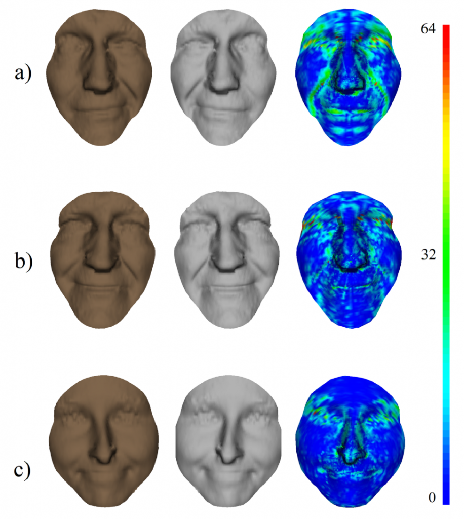
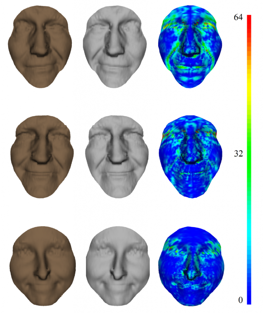
Scans of a smiling face: a) pre-operation, b) post-operation, c) control face.
Time: an essential fourth dimension in analysis
This technology is particularly well-suited to studying facial paralysis, as it takes time into account, and therefore the face’s dynamics. Dynamic analysis provides additional information. “When we look at a photo, it is sometimes impossible to detect facial paralysis. The face moves in three dimensions, and the paralysis is revealed with movement”, explains Mohamed Daoudi.
The researcher uses non-invasive technology to model the dynamics: a structured-light scanner. How does it work? A grid of light stripes is projected onto the face. This gives a face in 3D, depicted by a cloud of around 20,000 dots. Next, a sequence of images of the face making facial expressions is recorded at 15 images per second. The frames are then studied using an algorithm which calculates the deformation observed in each dot. The two sides of the face are then superimposed for comparison.
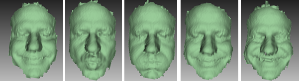
Series of facial expressions made during the scan.
Making 4D technology more readily available
Until present, this 4D imaging technique has been tested on a small number of patients between 16 and 70 years old. They have all tolerated it well. Doctors have also been satisfied with the results. They are now looking at having the technology statistically validated, in order to develop it on a larger scale. However, the equipment required is expensive. It also requires substantial human resources to acquire the images and the resulting analyses.
For Mohamed Daoudi, the project’s future lies in simplifying the technology with low-cost 3D capture systems, but other perspectives could also prove interesting. “Only one medical service in the Hauts-de-France region offers this approach, and many people come from afar to use it. In the future, we could imagine remote treatment, where all you would need is a computer and a tool like the Kinect. Another interesting market would be smartphones. Depth cameras which provide 3D images are beginning to appear on these devices, as well as tablets. Although the image quality is not yet optimal, I am sure it will improve quickly. This type of technology would be a good way of making the technology we developed more accessible”.
[1] Mohamed Daoudi is head of the 3D SAM team at the CRIStAL laboratory (UMR 9189). CRIStAL (Research center in Computer Science, Signal and Automatic Control of Lille) is a laboratory (UMR 9189) of the National Center for Scientific Research, University Lille 1 and Centrale Lille in partnership with University Lille 3, Inria and Institut Mines-Télécom (IMT).
[2] This project was supported by Fondation de l’avenir


Leave a Reply
Want to join the discussion?Feel free to contribute!