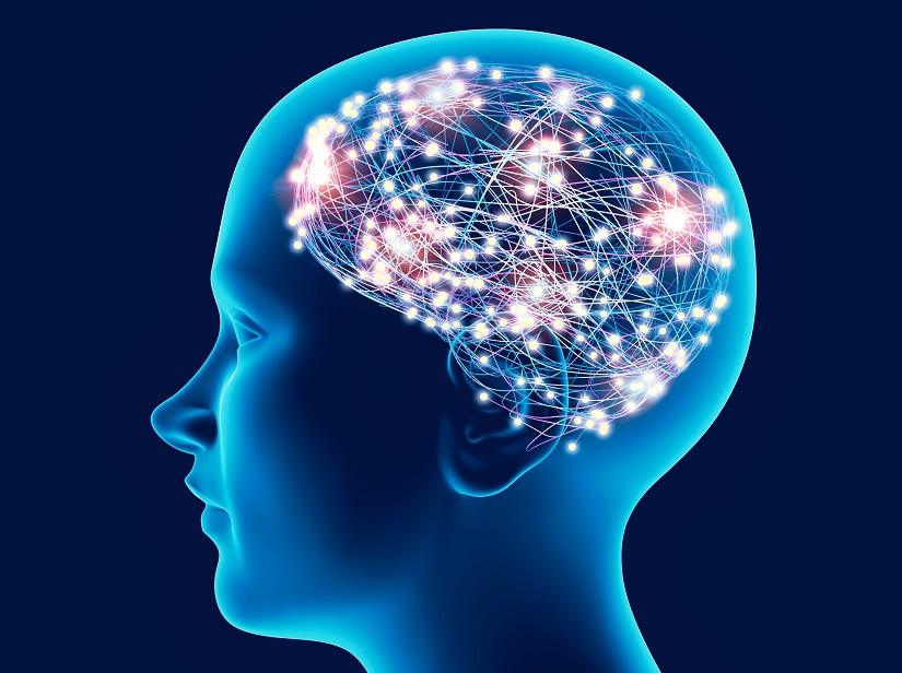
To observe what is going on in our brains, electroencephalography (EEG) is the tool of choice. By recording electrical activity generated by the brain, using electrodes placed on the scalp, it provides a wealth of information using a non-invasive technique. However, there are a number of problems with EEG, such as the fact that the skull limits the reading of signals emitted by the brain. This makes it difficult to obtain highly precise information on functional cerebral activity.
In an attempt to overcome these limitations, CEREBRO, a Horizon Europe project involving five European partners, was launched in 2022 for a 4-year period. Adrien Merlini, researcher in the Microwaves Department at IMT Atlantique (a CEREBRO partner), explains the aim of the project: “The idea is to inject a contrast medium that will, in theory, change the nature of the signals measured by the new EEG. The signals would then be more legible and offer more information from the brain, but still in a non-invasive way, i.e. without requiring surgery”.
The CEREBRO project is therefore looking for a way to reconstruct the entirety of measurable signals generated by brain activity, without having to open the patient’s skull. “This would unlock new medical and research possibilities with data that is not currently accessible,” says the researcher. This new data is mainly spatial and temporal. In other words, the better the signal captured by the EEG, the easier it is to know where brain activity is taking place, like a magnifying glass with better magnification. And it is also easier to track this activity over time, as with a screen that refreshes more frequently. Just as a videographer produces better images with a camera that can zoom in more and film faster, practitioners see the brain better with a tool that offers better spatial and temporal resolution.
The first contrast medium to explore the entire brain
For François Rousseau, data science researcher at IMT Atlantique, CEREBRO could be useful for clinical applications requiring a precise tool to measure brain activity, for example. “It could also be used for diagnosis and research on the brain, which remains difficult to understand.” Will this contrast medium soon be used in all EEGs? “It’s not an external measurement tool, as it requires the injection of a product into the body. So it’s going to be some time before it’s ready for validation and possible testing,” says the researcher.
To achieve the project’s objectives, the first pillar is therefore the development of the all-important contrast medium. As with all contrast mediums, the principle is based on highlighting areas of interest. In chemistry or biology, tracers can be used to track the spread of a product, by making it fluorescent or colored, for example, and therefore more visible than the rest of the medium. In the case of CEREBRO, the contrast medium affects electrical activity, which is detected during the EEG and provides the observer (e.g. a doctor) with information on the precise area in the brain where activity is taking place.
Understanding the benefits and limitations of EEG
There are two main types of brain imaging: anatomical and electrical. On the one hand, anatomical imaging enables us to characterize the tissues that make up the brain. This is the case with MRI (magnetic resonance imaging), which provides excellent spatial resolution with an accurate physical representation of tissues. The problem is that it does not provide enough information about brain activity.
On the other hand, electrical imaging of the brain using EEG shows what happens during a task or at rest. It offers good temporal resolution, but not spatial – hence the importance of the CEREBRO project. Activity in the brain is intense and needs to be recorded in relatively few measurements. “High-resolution research EEGs use nearly 256 electrodes to reconstruct potentially thousands of data points,” says Adrien Merlini. This technique provides sufficient information for certain applications, without being invasive.
That said, when EEG data is not sufficient to understand a patient’s condition, practitioners can go further. They then open the skull and insert electrodes directly into it, reducing signal attenuation caused by bone mass. However, the benefits can be marginal, and sometimes do not make up for the various costs (emotional, economic, temporal) of such an operation. This is where the CEREBRO project comes into its own, bringing the benefits of better EEG observation, without the need for major surgery on the patient.
CEREBRO: a European Pathfinder project
CEREBRO is one of the European Innovation Council’s Pathfinder projects, which focus on innovative and potentially risky factors. This type of project is typically experimental and can only be verified after testing. “IMT Atlantique is contributing to the modeling and reconstruction of source imagery,” says François Rousseau. Adrien Merlini emphasizes the importance of the initial phase of the CEREBRO project. “If, at the end of this process, we are able to demonstrate that the contrast medium we are aiming for is possible, the concept will be validated and we can move on to the next phase. The closer CEREBRO is to achieving this, the larger the project budget will be.”
CEREBRO is a long-term project which, if successful, will have a fundamental impact in many areas. It is in the initial phase of modality development to demonstrate proof of concept. In other words, even if the project succeeds at the end of the allocated years, it will have to be redesigned to take it to the next level. This offers promising prospects for learning more about the brain’s functional activity.


